About
Research Highlights
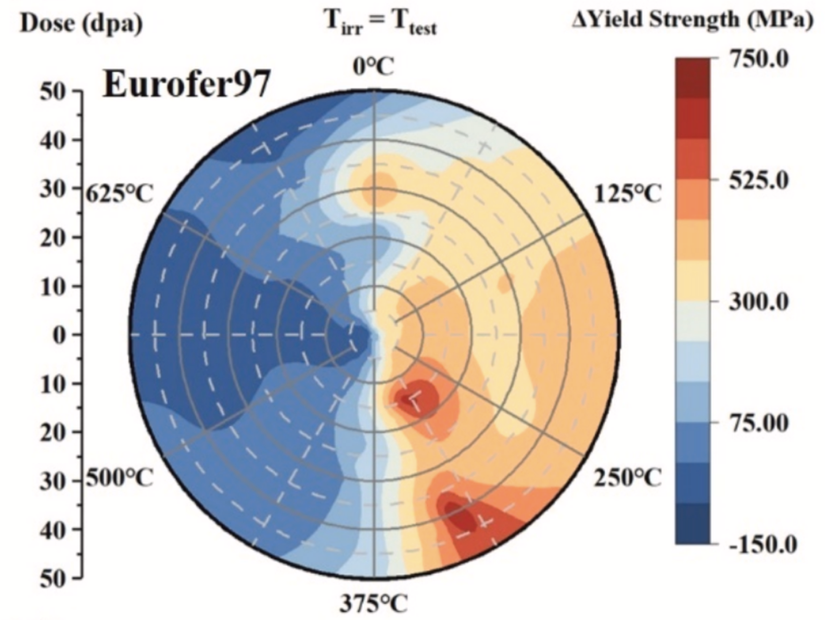
Radiation Hardening Effects on Eurofer97 Steel
Advanced machine learning analysis of radiation hardening in reduced-activation ferritic/martensitic steels. Comput. Mater. Sci. 251 (2025) 113773
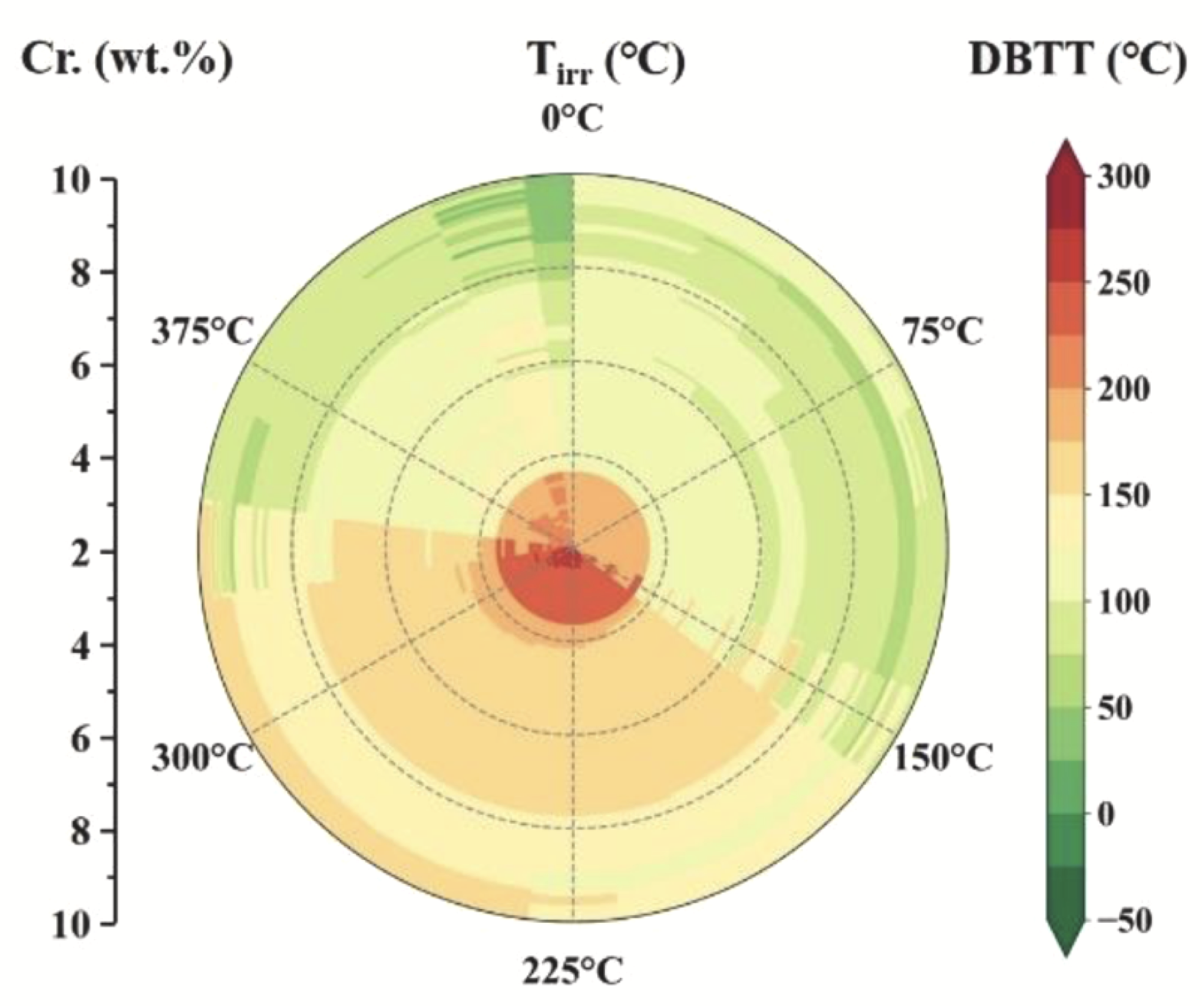
Effect of Cr on DBTT in Eurofer97 steels across different temperatures
A Data-driven machine learning model for radiation-induced DBTT shifts in RAFM steels. J. Nucl. Mater. 615 (2025) 155984
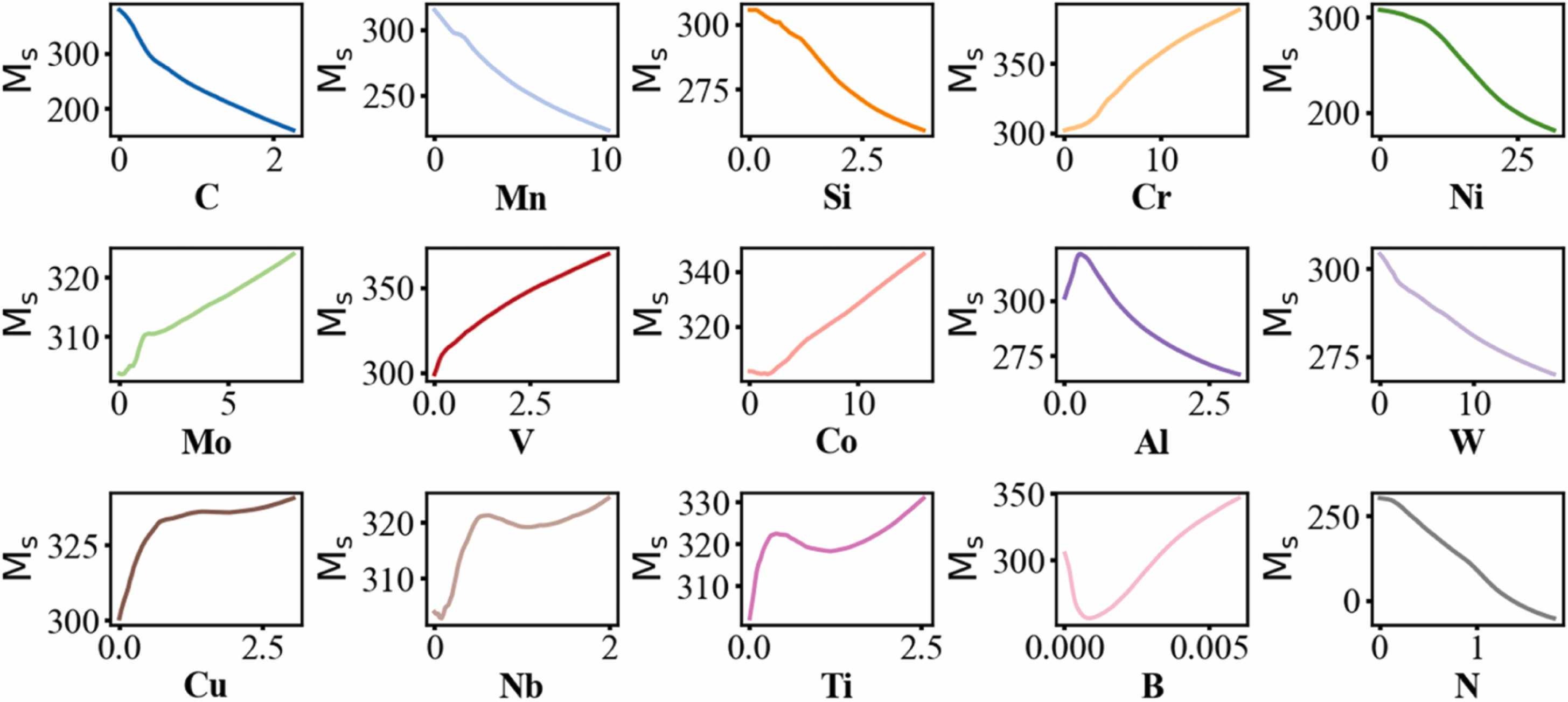
Sensitivity analysis showing how variations in individual alloying element concentrations (wt%) influence the predicted Ms.
Physically- and Knowledge-Informed deep learning for robust prediction of martensite start temperature in steels. Mater. Today Commun. 49 (2025) 113743
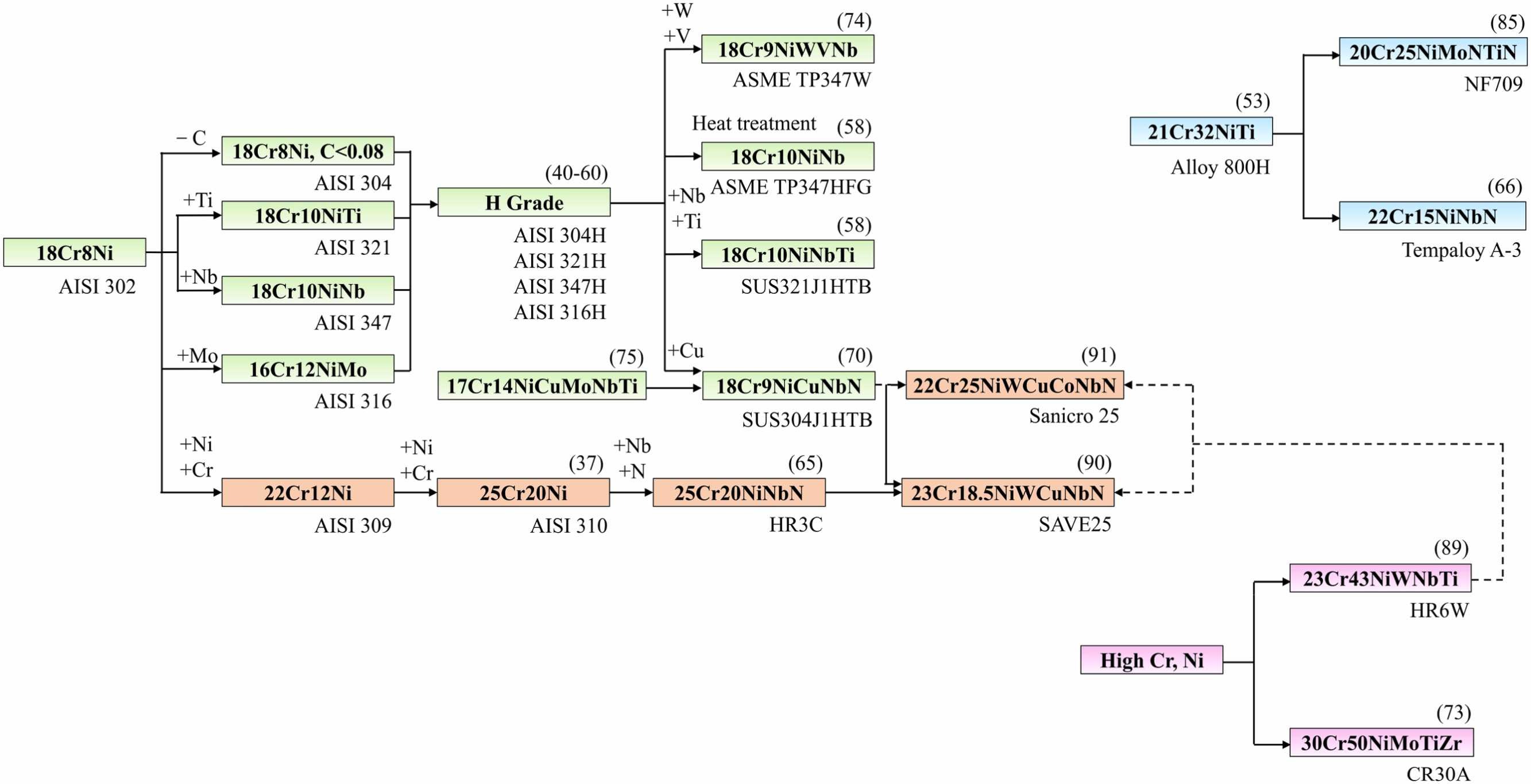
Development progress of austenitic steels, the numbers in parentheses indicate the creep rupture strength at 700°C for 100,000 h
A review on thermal expansion control in Fe-Ni-Cr austenitic alloys: From Invar effect to advanced power systems. Mater. Today Commun. 50C (2025) 114365
Ph.D. Thesis Resources
Materials & Processes
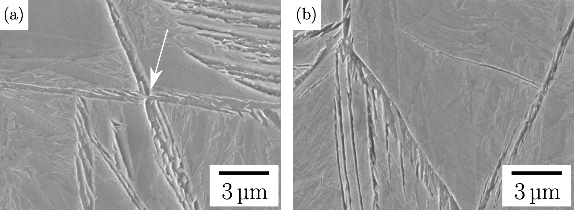
SEM of partial bainitic transformation, formed after transformation at 250°C for 10 h (a) with NiAl. The arrow indicates the intragranular nucleation of bainite from the NiAl precipitates. (b) Without NiAl, nucleation is from the austenite grain boundaries.
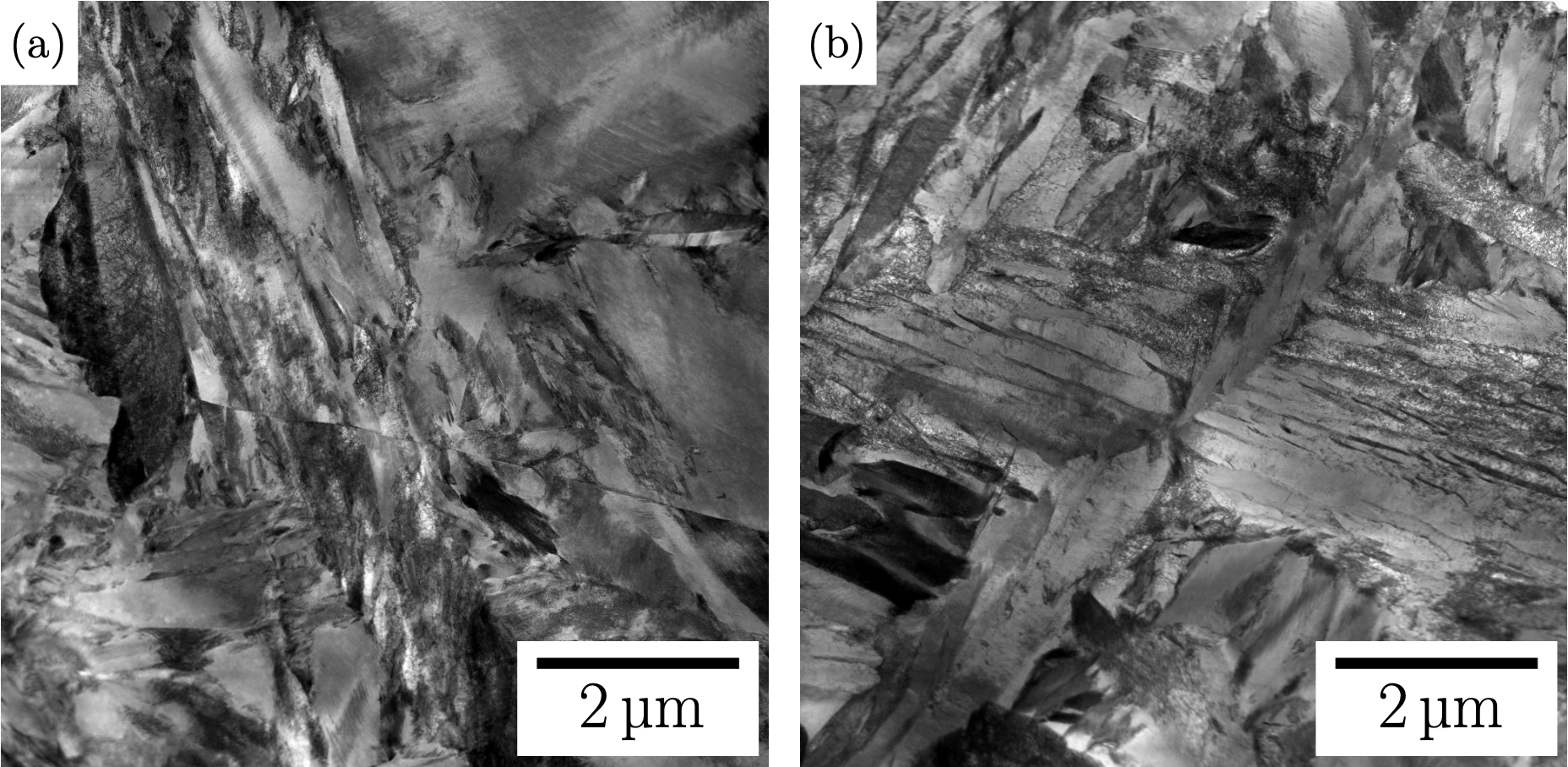
STEM bright field images of the transformed alloy. Microstructure of an interrupted bainitic transformation at 250°C for (a) 10 h. (b) 50 h, (b) shows the the nucleation of bainite from the NiAl precipitates where the bainite is seen to grow into many directions.
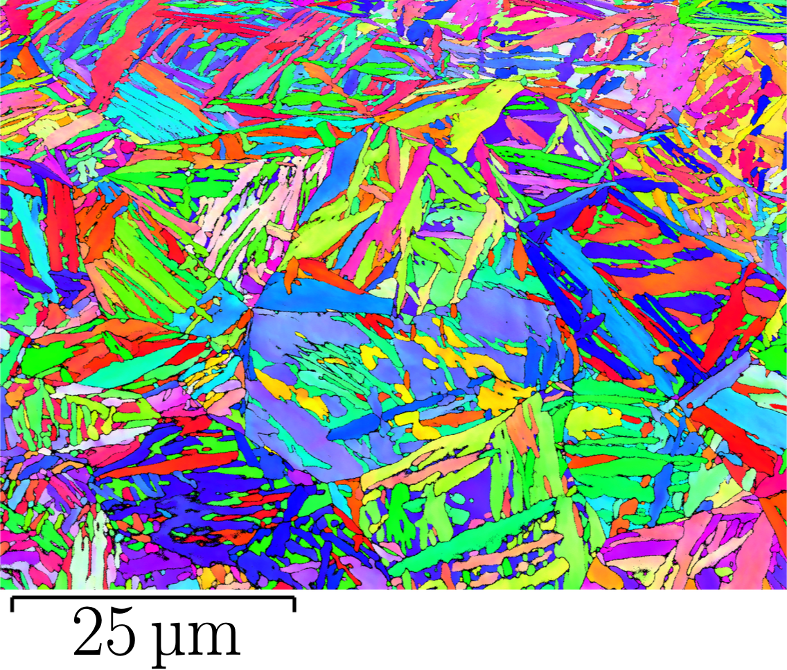
EBSD orientation image, sample transformed isothermally at 250°C for 150 h followed by cooling to ambient temperature. Bainite nucleated from NiAl promotes a more chaotic and finer bainite structure.
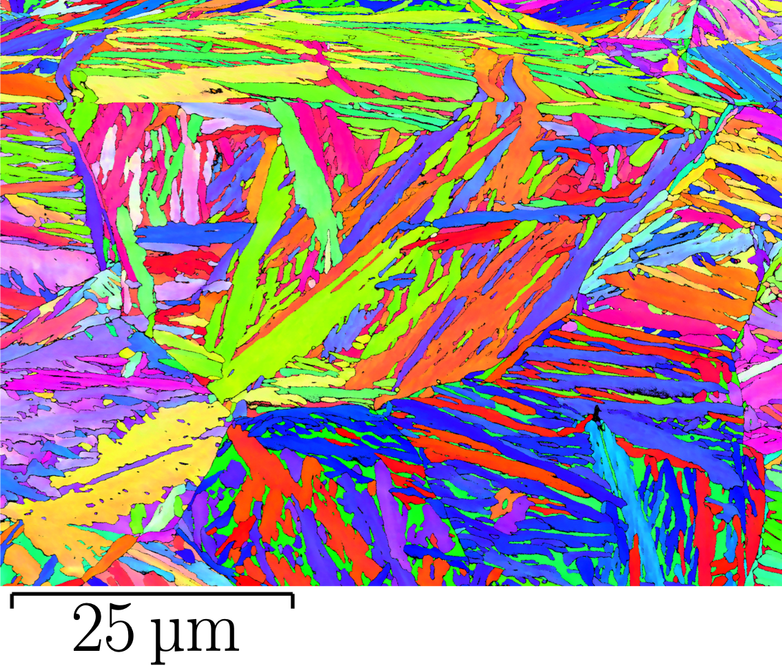
EBSD orientation image, sample transformed isothermally at 250°C for 150 h followed by cooling to ambient temperature. Grain boundary nucleated bainite.
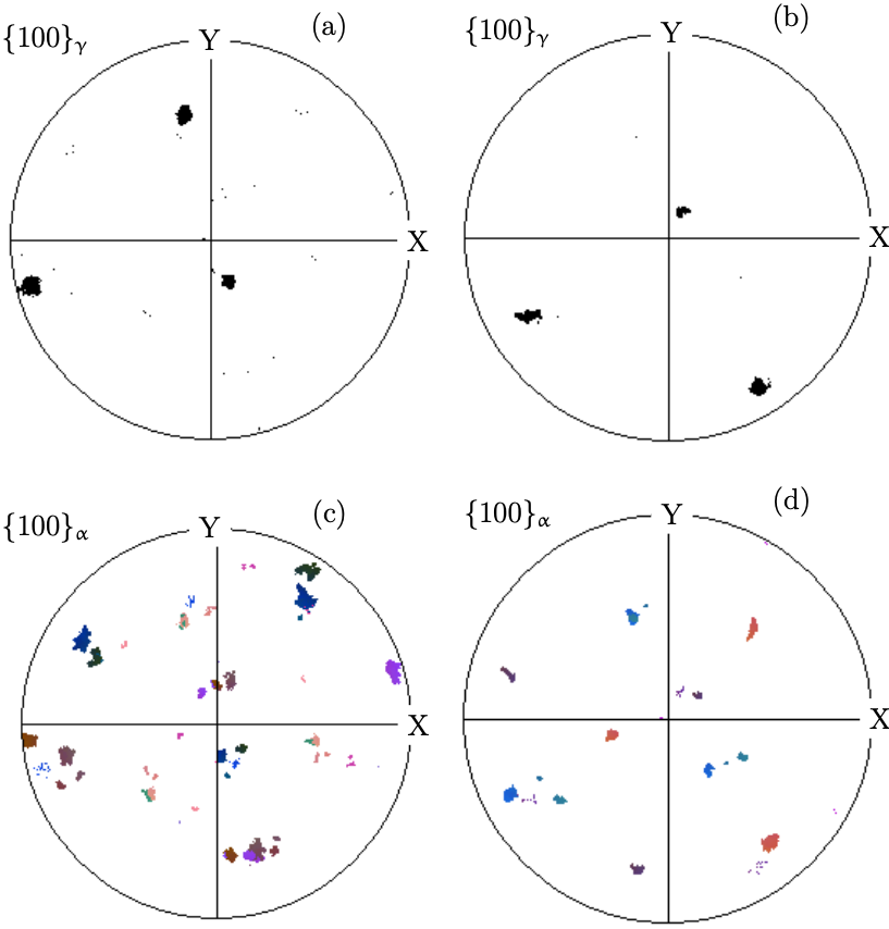
Pole figures from a parent austenite grain, (a and b) {100}γ pole figure, (c and d) corresponding {100}α pole figure, more variants within the austenite grain (c).
Explanation for Video 1
Explanation for Video 2